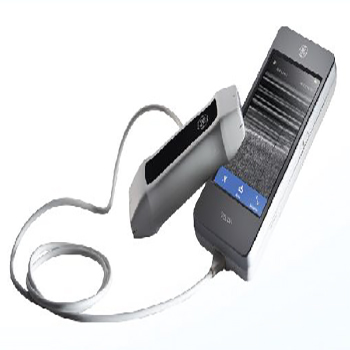
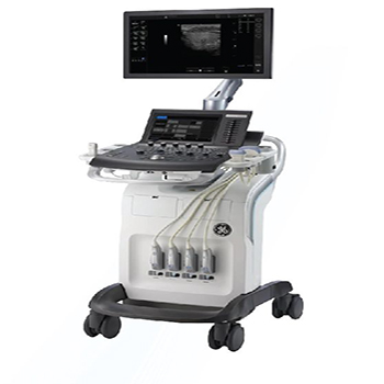
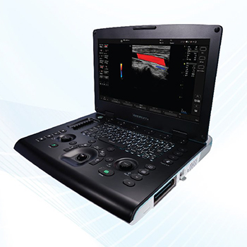
Point of Care Ultrasound Ultrasound Imaging Menu Cardiovascular Ultrasound General Imaging Ultrasound Women’s Health Ultrasound Primary Care Ultrasound Poiint of Care Uultrasound Venue 40 (Portable)-surpnsmgly Easy To use PDI highlights vascular sensitivityPDI quantification far diagnosis and monitoringChoose from three probes – always connected – far quick access.pre-configured application settingsIntuitive touch interfaceRespond fast with quick boot-upAdjustable height and screen Logiq e (Porta ble)-see Clearly. See Quickly. Guide Precisely. Optimized for head and neck. Permitting right-out-of-the-box imaging.Probe versatility – use a single probe for many applicationsSimple documentation; one click sends images to multiple storage locationsCompact and battery-operated. easily move from patient to patientLOGIQ e pre-sets help simplify the selection of the right system settings for each applicationPrecise visualization of law and slow blood flaw and accurate imaging of vascularity Vscan Access (Handheld)-Assessing Risk. Expanding Reach. Intuitive touchscreenGesture-based controlClinical protocols and automated controlsDamage-resistant screenDrop-tested dust-proof exteriorBattery-poweredLightweight for portabilityScan Coach tool to help optimize scan plane and probe positionGrowth tracking software to display foetal development over timeBluetooth® for wireless data transfer and m Health applications
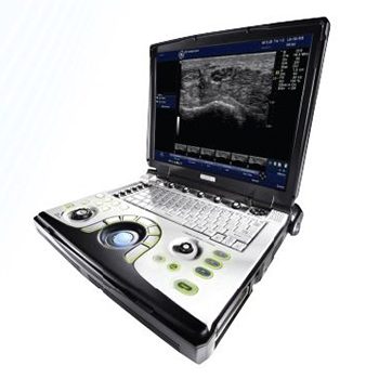
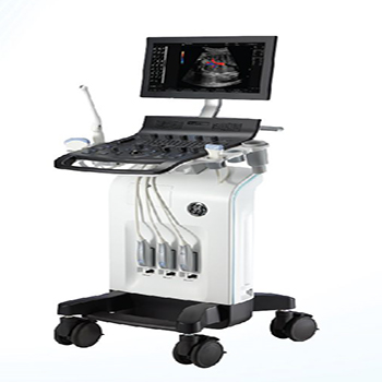
Primary Care Ultrasound Ultrasound Imaging Menu Cardiovascular Ultrasound General Imaging Ultrasound Women’s Health Ultrasound Primary Care Ultrasound Poiint of Care Uultrasound Versana Balance-care with canfidence 21.5″ LED Display .CrossXBeam and SRI-HD to define structure borders clearly.Height-adjustable console .Broad range of probes.Multiple ports to support a wide variety of exams.Make automatic 2D measurements.Scan Coach; 3D animation. illustrations and reference images to find the correct scan plane.Voice comments; capture recorded voice comments overlaid on images.Myocardial Doppler for imaging with colour overlay on the tissue image. Versana Premier-Powerful. Versatile. Productive. 21.5″ LED Display with 9.9″ touch screen.Height adjustable console.Gel wormer.Wizz function for easy image quality optimization.Needle Recognition clarifies the precise location of the needle point.Tricefy Uplink; send images to the Tricefy cloud wirelessly for consultation.TruScan to review and analyse images.Myocardial Doppler for imaging with colour overlay on the tissue image.Tomographic Ultrasound Imaging (TUI) to assess slices within a volume. Versana Active (Portable)-Advanced. Capable. Adaptable. Lightweight (5 KG with built in removable battery).New image processor with faster frame rates and updated algorithms.Follow-Up Tool to compare the current and prior exams side-by-side.Wizz function for easy image quality optimization.Scan Assistant- lets you create standardized exam protocols.Scan Coach – contextual reference tool for scan plane acquisition. Versa no Essentia I-Easy To use. Easy To Own. Automated measurement af IMTWizz function far easy image quality optimizationSmall footprint and lightweightSimple and fast to use with automated calculations3″ 16:9 LED Monitor3 active probe connectorsColor DopplerScan Coach – real-time reference information ta help locate the correct scan plane
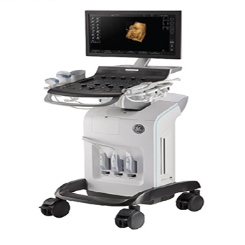
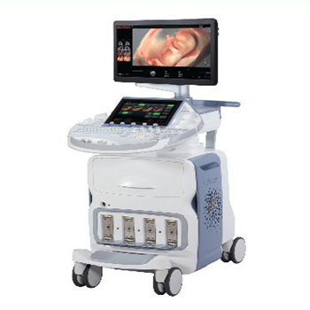
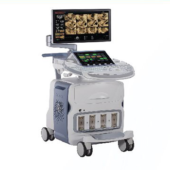
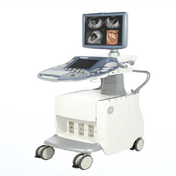
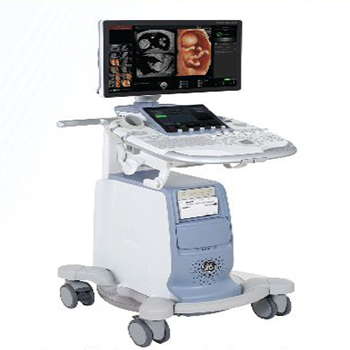
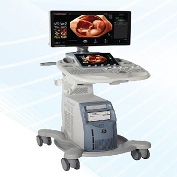
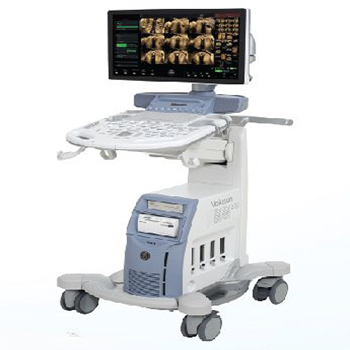
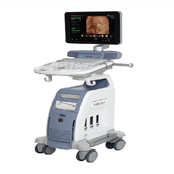
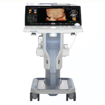
Women’s Health Ultrasound Ultrasound Imaging Menu Cardiovascular Ultrasound General Imaging Ultrasound Women’s Health Ultrasound Primary Care Ultrasound Poiint of Care Uultrasound Voluson E10 Radiantflow; delivers easy. fast visualization of even the tiniest of vessels.SlowflowHD for blood perfusion visualizations.HDlive™ technology suite helps easily obtain volume images with unprecedented depth and clarity.HDRes elevates tissue differentiation. border definition and fine resolution eSTIC. e4D. eSnapshot to optimize Electronic 4D.SonoCNS helps properly align and display views and measurements of the fetal brain.XDclear™ probe technology for achieving exceptional tissue and detail resolution. Voluson E8 Radiantflow; delivers easy. fast visualization of even the tiniest of vessels.SlowflowHD for blood perfusion visualizations.Radiance System Architecture for simplified scanning and foster processing speeds.Edison – Artificial Intelligence reduces keystrokes by 80% for foetal brain exams.proven interface coupled with automation and ergonomic design.4DView allows you to optimize. Manipulate and analyze volume ultrasound data offline. Voluson E6 SonoRenderlive facilitates 3D and 4D renderingXDclear™ probe technology for achieving exceptional tissue and detail resolutionUterine Trace for easy documentation of uterine shapeHDlive™ technology suite helps easily obtain volume images with unprecedented depth and claritySonoCNS helps properly align and display views and measurements of the foetal brain Voluson S10 Advanced VCI – Adjusts slice thickness to help enhance contrast resolution.Superb 2D and 3D/4D imaging. optimized for clarity and detail.Vibrant visualization of anatomy ond function with advanced color Doppler.HDlive™ technology suite; gets volume images with unprecedented depth and clarity.Consistent image quality even in the most difficult to image patients.SonoVCAD™heart for essential views of the foetal heart from a single STIC volume.3D Printing for rapid clinical prototyping. and parent bonding.Advanced VCI -adjusts slice thickness to help enhance contrast resolution. Voluson S8 Touch Excellent 2D and 3D/4D image qualityHDlive™ technology for unprecedented depth. clarity and exceptional anatomical realismVCI with OmniView to easily view irregularly shaped structuresSano-automation technologies to decrease complexity and increase exam consistencyVoluson xTouch; experience intuitive volume navigation on a 10.1″ touch panel Voluson S8-Extraordmary 23″ widescreen LED monitor.Report preview – provides access to completed measurements and trending Adaptive control panel.Battery Pack – provides up to 20 minutes of scan time.Superb 2D and 3D image quality optimized for clarity and detail.Color Doppler Quickly to quickly assess vascular anatomy and functions.Achieve the penetration needed for all exam and body types.HDlive™ technology for 3D and 4D imaging.HDlive™ technology for exceptional anatomical realism. Voluson P8 Excellent clarity and detail in 2D images.Expand clinical confidence with 3D/4D technology.Advanced Color Doppler for superb sensitivity into anatomy and function.Consistent imaging even in difficult-to-image patients.Sano-automation technologies.SonoRenderlive facilitates 3D and 4D rendering. Voluson SWIFT+ - 5″ High resolution full touch interface LED.Voluson core architecture. Light Weight and manoeuvrable.Sonolyst Improve quality and simplify fetal Anatomy Scans.Sano FHR for auto fetal heart rate measurement.Son biometry I HC. BPD. AC. FL. HL. CM Vp and cerebellum I Auto measurement.Uterine trace to obtain coronal plane of uterus.SonoCNS helps properly align and display views and measurements of the foetal brain.Battery Backup.
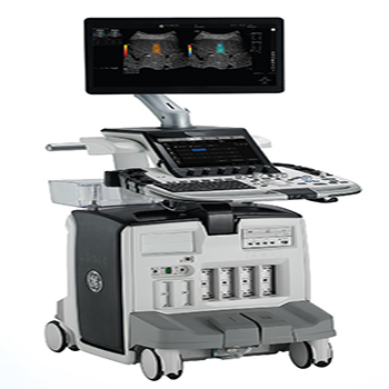
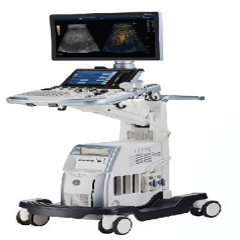
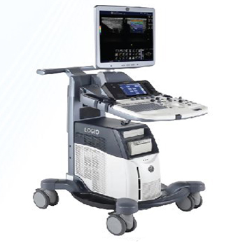
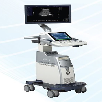
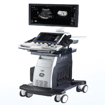
General Imaging Ultrasound Ultrasound Imaging Menu Cardiovascular Ultrasound General Imaging Ultrasound Women’s Health Ultrasound Primary Care Ultrasound Poiint of Care Uultrasound Logiq E10S 22″ OLEO screen12″ multi-touch screenUGAP, Micro-Vascular ImagingAuto Doppler AssistStart Assistant enables you to experience end-to-end workflowOB/Breost/Tyroid Tools for labelling, measuring and describing lesions, nodules, lymph nodes and parathyroidAcquire images in your preferred order or easily reorder images within the examB-Steer + to enable enhanced visualization of the needles structureMulti-dimensional 2D sharewove Elostogrophy“Any-Plane” function for 3D and 4D data Logiq S8 23″ LED screen10.1″ multi touch screenS-Agile Architecture; dynamic image acquisition for all body typesE-Series Transducers for improved sensitivity, penetration and image uniformityHigh-Definition Speckle Reduction Imaging (SRI-HD)CrossX Beam; combines multiple images into one clear appearanceCoded Harmonic Imaging for enhanced near field resolution and far field penetrationLOGIQ View facilitates excellent visualization and more clinical information.B-Flow Imaging enables direct visualization of blood flow, without the issues of doppler Logiq S7 23″ LED screen10.1″ multi touch screenS-Agile Architecture; dynamic image acquisition for all body typesE-Series Transducers for improved sensitivity, penetration and image uniformityHigh-Definition Speckle Reduction Imaging (SRI-HD)CrossX Beam; combines multiple images into one clear appearanceCoded Harmonic Imaging for enhanced near field resolution and far field penetrationLOGIQ View facilitates excellent visualization and more clinical information.B-Flow Imaging enables direct visualization of blood flow, without the issues of doppler Logiq P7 Large 21.5″ monitor and accessible 10.4″ touchscreenTouch Control; easily adjust imaging parameters on touch panelPhoto Assistant App; combine onotamicol photos and images in same report2D Shear Wove Elostography – Quantitative estimate of tissue elasticityRemote Control App – Operate the system from on Android phone or tabletMy Trainer softwareCRl,SRI, ATO, AO Logiq P9 Large 21.5″ monitor and accessible 10.4″ touchscreenTouch Control; easily adjust imaging parameters on touch panelPhoto Assistant App; combine anatomical photos and images in same reportHD Color – Sensitivity for visualizing small vessels and slow flow2D Shear Wove Elostography – Quantitative estimate of tissue elasticityRemote Control App – Operate the system from on Android phone or tabletSonoDefense – Powerful data security features to help guard against costly breachesCRl,SRI, ATO, AO
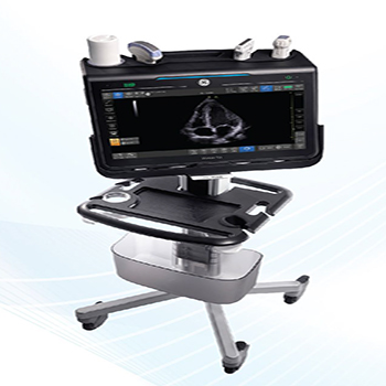
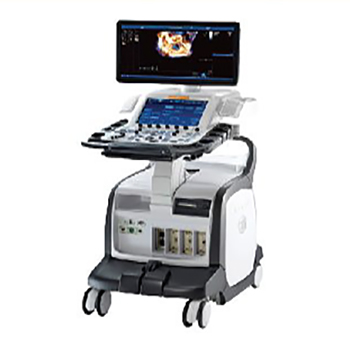
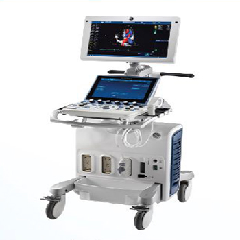
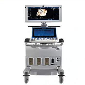
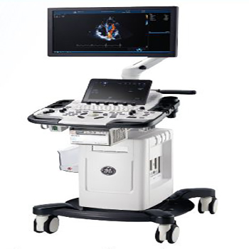
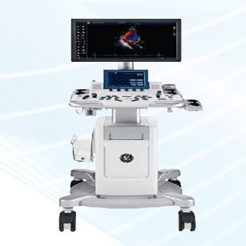
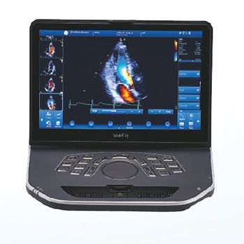
Cardiovascular Ultrasound Ultrasound Imaging Menu Cardiovascular Ultrasound General Imaging Ultrasound Women’s Health Ultrasound Primary Care Ultrasound Poiint of Care Uultrasound Vivid E95 High resolution 24′ wide LED monitorAFI ILV, RV, LA)40 MVQ, AVQ, LVQ, RVQ, TVQAuto measurement 2DAuto Spectrum RecognitionAuto VQ, Flexi LightHD Colour, 40 MarkerAnatomical M-mode both on line & offline Vivid S60 21.5″ wide screen HD display12″ multi-touch LCD screen with tablet like performanceAF! ILV, RV, LA}Al Auto measurement 2DStress echo, TVI, TOI, Tl TSIFlex Fit – Adjust control panel and monitorSmart standby without AC power Vivid S70 21.5″ wide screen HD displayErgonomic FlexFit designAuto measurement 2DAFI, Stress echo, TVI, TOI, TT. TSIAuto Spectrum Recognition12″ ultra-high-resolution wide screen format4D TEE Capabilities Smart standby Vivid T9 21.5″ wide screen HD displayErgonomic FlexFit designIntuitive control layout includes the 10.Uil multi-touch screenCardiac auto doppler- artificial intelligence-based toolAutoEF 2.0 – second-generation tool for assessing and quantifying ventricular wall motionStress EchoAFI, Tl TVI, TOI. TSI, Q-AnalysisAuto measure 20Articulating arm Vivid T8 21.5″ wide screen HD displayIntuitive control layout includes the 10.Uil multi-touch screenLVO Contrast, Tissue Tracking, Adv. Qscan Imaging, Q-AnalysisScan Coach; provides reminders/refresh informationSmart StandbyTissue Velocity Imaging – captures dynamic information from moving heart tissueBlood Flow ImagingStress Echo Vivid iq Light weight 5.2kgAl Auto measurement 2DAFI ILV, RV, LA}Stress EchoAuto EF, TT. TVI, TOI, TSI. Q-AnalysisAuto Spectrum RecognitionClosed surface design – allows cleaning with disinfectant solutions